MOBILE
Monitoring of Bone Impact Levels at Exeter
MOBILE is led by Dr Vicky Stiles and Dr Matthew Ellison to drive forward research and innovation in bone health.
Mission
MOBILE (Monitoring of Bone Impact Levels at Exeter) is an interdisciplinary group committed to advancing research, tools and technologies that promote bone health across the life course.
Our mission is to develop better research methods to precisely monitor bone-specific activity and to co-develop research-informed, user-facing tools that measure bone-specific impact activity, enhance skeletal health and help prevent osteoporosis and injury. We work through equitable partnerships, combining expertise in biomechanics, digital health innovation, participatory research, data science and statistics and commercialisation to deliver real-world impact.
Aims & Expertise
- Designing, validating and applying innovative methods to monitor bone-specific mechanical loading
- Increasing understanding of how physical activity influences bone strength and skeletal health across different populations
- Co-developing and evaluating digital health solutions in collaboration with end users, health systems and industry
- Informing public health strategies for osteoporosis prevention and management
- Fostering interdisciplinary training, mentoring, and capacity building in bone health, digital health and innovation
.jpg)
Group Members
Zisheng Xu (PhD Student)
Meshary Algadheb (PhD Student)
Mia Pollington
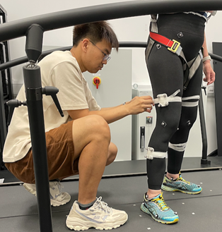
This video demonstrates the collection of biomechanical data using Vicon, where markers (spheres) are placed on the lower limb to track movement during hopping. The data is then combined with musculoskeletal modeling in OpenSim to estimate bone loading.
Developing bone-specific activity metrics
We design and refine methods to quantify bone-loading characteristics of everyday physical activity. This includes identifying movement patterns with osteogenic potential and relating estimated or measured mechanical loads to specific types of activity in different populations. Our aim is to create metrics that reflect real-world bone-relevant activity and support interventions across the lifespan.
Estimating bone loads using wearable sensors
We develop and validate methods to estimate mechanical loading at key skeletal sites using accelerometry and other wearable sensor data. Our work involves modelling relationships between sensor outputs (e.g. peak acceleration) and biomechanical loading metrics (e.g. peak ground reaction force, loading rate), and testing sensor placements and algorithms in diverse populations. This allows us to quantify the bone-loading potential of everyday movements outside laboratory settings.
Using musculoskeletal modelling to estimate loading on the bone
We combine biomechanical data and imaging techniques, to develop, evaluate and apply musculoskeletal models that allow us to better understand how footwear, exercise and training affects loading on the bone to either reduce the risk of injury or development of bone diseases.
Case Studies
Physical activity and bone health in pre- and post-menopausal women: UK Biobank
Methods: N~2500 women with cross-sectional measures of bone health (heel ultrasounds) and physical activity (1wk accelerometery). Multiple regression analyses - with incrementally increasing intensity thresholds - were carried out to determine the optimal impact required to benefit bone health.
Findings: We found that as little as 1-2min/day of physical activity of at least moderate-impact (≥750mg), was favourably associated with better bone health. We also demonstrated that a threshold commonly used in such studies – that of MVPA (≥250mg) - was not associated with bone health. (Stiles 2017)
Physical Activity and Bone Health in Children
.jpg)
This study explored how physical activity supports bone development in children aged 11-12 using novel analysis of accelerometer data. Traditional methods often miss the short bursts of high-impact movement that benefit growing bones. By analysing raw, wrist-worn data in 1-second intervals and fine-grained intensity bands, researchers found that just 10 minutes per day of high-intensity activity (above 700 mg), about the intensity of running at 10 km/h, was linked to stronger, denser bones in children. These findings provide a more accurate way to assess bone-relevant activity and clarify the dose needed to build peak bone mass during childhood.
Developing wearables to monitor loads most relevant to bone health
Project lead: Dr Vicky Stiles
An improved ability to objectively monitor simple, meaningful characteristics of external training load on a large population scale will enhance our understanding of the influence of training load on injury and performance in athletes.
Working with Activinsights Ltd and using wrist-worn activity monitors (wearables; accelerometers), we have generated new training load metrics from raw acceleration data to allow training programmes for runners to be monitored 24-7. Capturing characteristics of training, rest and recovery are important for enhancing performance and preventing injury. Similar techniques are also being developed for tennis players and other populations engaging in high-performance activity.
Modelling the osteogenic potential of different exercises on the femur
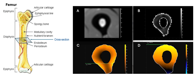
We are examining how different physical activities influence mechanical stress and therefore osteogenic potential on the femur. We are also investigating how the osteogenic response may vary when we alter bone structural properties (e.g., Young’s Modulus and Poisson’s Ratio) to simulate both healthy and osteoporotic bone conditions.
Cross-sectional slices of the femur showing the ring of cortical bone surrounding the medullary canal are presented as follows:
A) Single MRI slice
B) Segmented cortical bone
C) Horizontal shear stresses on the cortical bone (yellow = higher stress)
D) Axial stresses (in the direction of loading) on the cortical bone (yellow = higher stress)
Modelling bone stresses on the metatarsal during running
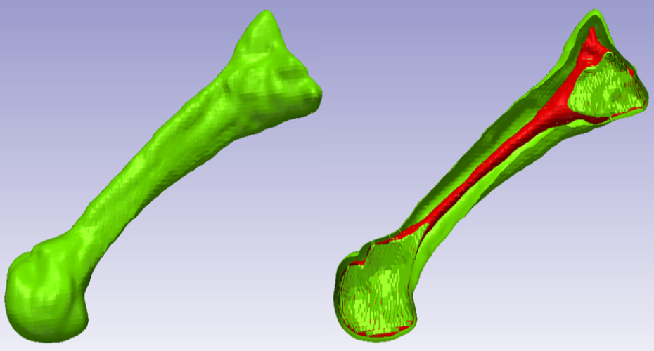
Project lead: Dr Matthew Ellison
In order to understand bone injuries in runners and military populations we need to measure the stress the bone experiences during each step.
We are working with Engineering to construct a 3D representation of the bones in the foot and lower leg from medical images.
We measure each person’s running style in our gait laboratory and then calculate how much stress the bone experiences during each step. From this we can understand if running in different conditions, such as different footwear, changes the amount of stress acting on the bone.