Overview
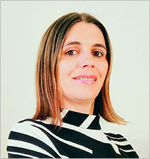
Liliana is a Diagnostic Radiographer and a lecturer in Medical Imaging, she joined to the University of Exeter in 2017.
Liliana finished her degree in Diagnostic Radiography in 2005 and started working as a Radiographer in Portugal. During 12 years she obtained clinical experience in General Radiography, Computed Tomography, Mammography, Densitometry, and Magnetic Resonance.
She had combined the clinical practice with teaching for 10 years as she was a part-time invited lecturer at School of Allied Health Sciences in Portugal between 2007 and 2017.
She finished her master degree in 2012 with a dissertation about “Contrast Medium Volume Optimization in Abdominal CT on Basis of Lean Body Weight” and she got the title of specialist lecturer in 2016 with a research about “Radiographer precision in bone densitometry”.
Liliana now combines teaching with her PhD, her research topic is “Micro and Macro structural predictors of osteoporotic vertebral fractures”.
Qualifications
- 2020 - Fellow of the Higher Education Academy (Recognition reference: PR179518)
- 2018 - PhD candidate, Research project: “Micro and Macro structural predictors of osteoporotic vertebral fractures”.
- 2012 - MSc in Medical Informatics at Faculty of Medicine in University of Porto, Portugal.
- 2005 - BSc in Radiology (Diagnostic Radiography) at School of Allied Health Sciences in Polytechnic of Porto, Portugal.
Research
Research interests
Liliana is also currently undertaking a PhD, looking at Micro and Macro structural predictors of osteoporotic vertebral fractures.
Liliana's research interests are mainly Osteoporosis, Bone densitometry, Contrast Media and Computed Tomography.
Publications
Key publications | Publications by category | Publications by year
Publications by category
Journal articles
Ferreira Santos P, Rodrigues L (2016). Eagle’s syndrome - a case report. international journal of stomatology & occlusion medicine, 8(S1), 54-59.
Rodrigues L, Sampaio R, Coimbra M (2013). Contrast Medium Volume Optimization in Abdominal CT on Basis of Lean Body Weight.
American Journal of Biomedical Engineering,
3, 22-26.
Abstract:
Contrast Medium Volume Optimization in Abdominal CT on Basis of Lean Body Weight
Computed tomography (CT) in abdominal imaging has undergone great advances in recent years. The administration of iodinated contrast media (CM) has evolved along with the evolution of the CT equipment. The most common method for calculation of the CM volume in abdominal CT is based on total body weight (TBW), but in this way some patients get a lower dose and some other patients get a higher dose that the optimal dose they need. In the literature there are many formulas to calculate the CM volume to be administered in abdominal CT, but practice shows that the variability of enhancement is wide. The main objective of this study is to verify that the volume of intravenous CM in abdominal CT calculated on basis of lean body weight (LBW) allows good liver enhancement with small amplitude of variation around the mean, that is, with less variability in enhancement than CM volume calculated on basis of TBW. In conclusion the calculation of the volume of CM on basis of LBW translates into a low variability of liver enhancement–25.50 HU. In our results 96.05% of sample belongs to a good hepatic enhancement. If we stratify sample by groups of BMI, we also verify no significative differences between slims and fat patients when analysed mean hepatic enhancement.
Abstract.
Chapters
Rodrigues L (2013). Estudo Radiológico do membro superior: mão, punho e antebraço. Estudo da Idade Óssea. In Rua Ventura S (Ed) Técnicas e Procedimentos em Radiologia Convencional, Portugal: LIDEL Edições Técnicas, Lda, 38-63.
Publications by year
2016
Ferreira Santos P, Rodrigues L (2016). Eagle’s syndrome - a case report. international journal of stomatology & occlusion medicine, 8(S1), 54-59.
2013
Rodrigues L, Sampaio R, Coimbra M (2013). Contrast Medium Volume Optimization in Abdominal CT on Basis of Lean Body Weight.
American Journal of Biomedical Engineering,
3, 22-26.
Abstract:
Contrast Medium Volume Optimization in Abdominal CT on Basis of Lean Body Weight
Computed tomography (CT) in abdominal imaging has undergone great advances in recent years. The administration of iodinated contrast media (CM) has evolved along with the evolution of the CT equipment. The most common method for calculation of the CM volume in abdominal CT is based on total body weight (TBW), but in this way some patients get a lower dose and some other patients get a higher dose that the optimal dose they need. In the literature there are many formulas to calculate the CM volume to be administered in abdominal CT, but practice shows that the variability of enhancement is wide. The main objective of this study is to verify that the volume of intravenous CM in abdominal CT calculated on basis of lean body weight (LBW) allows good liver enhancement with small amplitude of variation around the mean, that is, with less variability in enhancement than CM volume calculated on basis of TBW. In conclusion the calculation of the volume of CM on basis of LBW translates into a low variability of liver enhancement–25.50 HU. In our results 96.05% of sample belongs to a good hepatic enhancement. If we stratify sample by groups of BMI, we also verify no significative differences between slims and fat patients when analysed mean hepatic enhancement.
Abstract.
Rodrigues L (2013). Estudo Radiológico do membro superior: mão, punho e antebraço. Estudo da Idade Óssea. In Rua Ventura S (Ed) Técnicas e Procedimentos em Radiologia Convencional, Portugal: LIDEL Edições Técnicas, Lda, 38-63.
Liliana_Rodrigues Details from cache as at 2023-03-20 18:23:10
Refresh publications
External Engagement and Impact
Committee/panel activities
Memberships:
- Society and College of Radiographers - 27442
- HCPC Registered Radiographer – RA77658
- European Society of Radiology - 601710
- Radiographer professional title recognized by the Central Administration of the Health System (ACSS) – C-025362143
- National Osteoporosis Society – 718806-C
- ATARP (Portuguese Radiographers Association) - 2569
Invited lectures
2011 November (26) to December (10) – Invited trainer of the professional training course titled "Multislice CT", promoted by the Union of Sciences and Health Technologies, 20 hours.